-
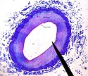
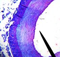
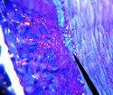
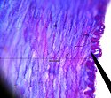
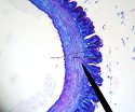
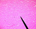
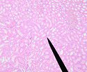
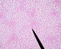
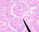
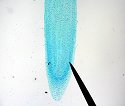
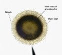
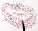
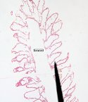
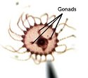
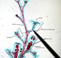
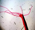
Histology PageHistology is the study of tissues. Below are images to help you review/study for the histology section of your exams. (Click to enlarge, some are labeled)!Arteries/VeinsArteries and veins have three basic layers (or tunics): the tunica intima, the tunica media, and the tunica adventitia. Each layer is composed of different types of tissue:-The tunica intima is the inner-most layer and is mainly composed of an inner lining of endothelium-The tunica media is the middle layer and is mainly composed of smooth muscle tissue-The tunica adventitia is the outer most layer and is mainly composed of elastic tissueThings to note:-Arterial walls are much thicker than veins due to the higher pressure found in arteries as compared to veins. Compare the different thicknesses of the different layers.ArteriesVeinsCardiac TissueCardiac tissue is striated but looks less organized. The cells are in a branched shape with intercallated disks in-between.Things to note:-Striations and incalated disksKidneyKidneys are the bodies osmoregulatory system. The filtering unit of the kidney is the nephron.Things to note:-The renal corpuscle (Bowmans capsule + glomerulus)-Proximal convoluted tubule: is generally smaller and more starshaped, also has cuboidal epidermal cells with microvilli (called a "brush border on the microscope")-Distal convoluted tubule: lumen is generally larger and more round than the PCT. Cuboidal cells are smaller, no bush border existsOnion Root Tip (Mitosis)Onion root tips are good tissues to look at when trying to see stages of mitosis. The growth of root occurs at the root tip so there are many cells undergoing mitosis. Below are images an onion root tip with the chromosomes, cell walls, and spindle fibers stained.Things to note:-Try to find all of the phases of mitosis in the following images and on your slides. Interphase is also very easy to find.-Compare how the genetic material in blue/purple looks between mitosis and interphase.-The cell walls and sometimes the spindle fibers are also visible in green.PoriferansCnidariansPlatyhelminthesNematodaAnnelida
Last Modified on March 21, 2025

